Kingston University
Kingston University
Jean-Christophe Nebel

Structure-based description of binding sites

Duration: 2003-2004

Jump to: Project description | Pilot Study | Keywords

Project description

The simultaneous alignment of several protein sequences is now an essential step in protein analysis. It allows the detection of distant homologues and the identification of patterns from which protein activities can be inferred.

With the increasing availability of protein 3D structures, the generation of biologically meaningful 3D patterns or 3D motifs from the simultaneous alignment of several protein structures becomes an exciting prospect.

Applications:

As a member of the Bioinformatics Research Centre (protein structure group) at the University of Glasgow, I investigated how techniques developed in 3D Vision and Graphics can be applied to the recognition of protein binding sites and protein docking when protein 3D structure is known.

  • Cavity detection (pattern recognition)

    • Voxelisation of the protein 3D space.
    • Each voxel is assigned an atom density.
    • Detect potential binding sites by comparing atom density of voxels with their neighbours'.

  • 3D geometrical alignment of cavity with known sites

    • Subdivide voxels using octrees around potential binding sites.
    • Filter by comparing volumes.
    • Align using principal axes.
    • Attempt to align using ICP registration (Interative Closest Point).

  • Generation of a 3-D chemical map of the potential binding site

    • Chemical properties are assigned to voxels defining the cavity (based on properties of amino acids they contain).

  • Comparison of chemical map of the cavity with maps of known sites

    • Generate a 2-D representation of topological arrangement of chemical properties (more manageable and maps are already geometrically aligned).
    • Compare graphs by computing Maximal common subgraphs.

    Pilot Study: Description of ATP binding sites

    In collaboration with: David Gilbert (Professor of Bioinformatics) and Pawel Herzyk (Biochemistry)

  • Cavity detection (case of crystalline profilin--actin, 2BTF.pdb)

    Our algorithm is based on:

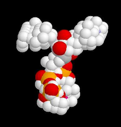
    Protein with ligand (ATP)
    Configuration of the ATP mollecule
    Residues involved in ATP binding (SITE field in PDB file)
    Binding site defined by CASTp
    Cavity defined by PASS
    Cavity defined by our algorithm

  • Binding Site Geometry (case of Tryptophanyl-TRNA Synthetase, 1MAW.pdb)

    Geometry is expressed by the volume of the cavity and the values of its principal axes.

    View of the protein composed of 6 identical chains, each of them binding to an ATP mollecule
    ATP mollecule configuration for chains A, B, C, D, E & F

    Shape variations (principal axes):
    12.5 5.6 1, 18.3 08.0 1, 17.2 8.4 1,
    10.0 5.8 1, 18.8 11.0 1, 21.0 9.8 1

    5 binding sites recognised by CASTp (volume +-10%)
    6 cavities recognised by our algorithm (volume +-13%)

    Shape variations (principal axes):
    3.3 2.4 1, 3.0 1.5 1, 4.6 3.1 1,
    3.7 2.2 1, 3.8 1.6 1, 5.7 2.6 1

    Research interest keywords

    Structural Bioinformatics, 3D Graphics, 3D Structure, Binding Site, Protein Docking


    Last updated in June 2006
    j.nebel@kingston.ac.uk