
|
|
|
Jean-Christophe Nebel
|
|
 |
CLAPA (Cleft Lip and Palate) and orthognathic surgery planning
Position: Consultant
Funding: Chief Scientist Office (CSO) of the Scottish Executive Health Department
Duration: 2001-2003
|
| Project description |
Orofacial clefting is the most common craniofacial birth defect, in Scotland the incidence of cleft lip and/or palate is approximately 1 in every 650 live births. The aim of the project is to address the standard of clinical and surgical care provided for those children who are affected.
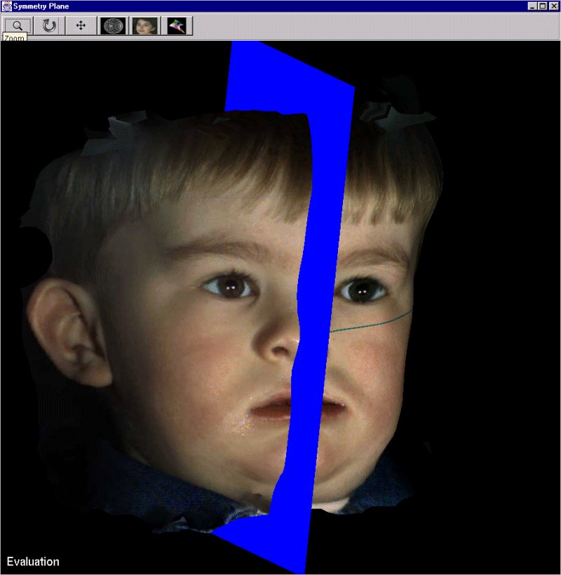
Our study aims to undertake advanced morphometric (quantitative shape analysis) assessment of non-cleft, cleft and surgically-managed cleft-patients in order to assess the influence of surgical lip repair on facial morphology. To achieve this goal, we access accurately the soft tissue morphology by using 3D imagers.
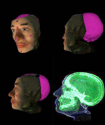
Over the last three decades orthognathic surgery has become a routine procedure for the correction of facial deformity. The most commonly used method of planning is to cut up profile photographs magnified to the same size as the standardized lateral skull radiograph. These are then superimposed over the cephalographs. The various portions of soft tissue and underlying bone are moved around to produce the most acceptable result. Unfortunately, radiographic and photographic registration and superimposition are inaccurate. The prediction of the postoperative appearance is limited to two dimensions. To address these problems a truly 3D modality of planning is required. Using a stereo photogrammetry machine, we are able to capture the 3D geometry of the soft tissue air boundary - that is a three dimensional shape of the face itself. The acquisition of 3D images of the underlying skeletal hard tissue is made using a CT scanner. We present the process we have developed which allows the registration of these 2 sets of 3D geometry. The registration itself is performed semi automatically using a ICP based software.

| Research interest keywords |
medical applications, 3D scanners
| Publications |